Category:Microscopic images of Allium cepa
Jump to navigation
Jump to search
Subcategories
This category has the following 2 subcategories, out of 2 total.
R
Media in category "Microscopic images of Allium cepa"
The following 57 files are in this category, out of 57 total.
-
 Allium cepa with KMnO4 and Phosphate.jpg 2,288 × 1,907; 816 KB
-
 Allium cepa-cell nucleus.jpg 403 × 444; 37 KB
-

-
 Cellule d'oignon en métaphase et anaphase.jpg 926 × 495; 82 KB
-
Chromosome-Dynamics-Visualized-with-an-Anti-Centromeric-Histone-H3-Antibody-in-Allium-pone.0051315.s003.ogv 8.0 s, 1,280 × 720; 1.53 MB
-
Chromosome-Dynamics-Visualized-with-an-Anti-Centromeric-Histone-H3-Antibody-in-Allium-pone.0051315.s004.ogv 7.8 s, 1,280 × 720; 1.9 MB
-
Chromosome-Dynamics-Visualized-with-an-Anti-Centromeric-Histone-H3-Antibody-in-Allium-pone.0051315.s006.ogv 8.0 s, 1,280 × 720; 2.12 MB
-
Chromosome-Dynamics-Visualized-with-an-Anti-Centromeric-Histone-H3-Antibody-in-Allium-pone.0051315.s007.ogv 8.0 s, 1,280 × 720; 2.1 MB
-
Chromosome-Dynamics-Visualized-with-an-Anti-Centromeric-Histone-H3-Antibody-in-Allium-pone.0051315.s008.ogv 8.0 s, 1,280 × 720; 2.75 MB
-
Chromosome-Dynamics-Visualized-with-an-Anti-Centromeric-Histone-H3-Antibody-in-Allium-pone.0051315.s009.ogv 8.0 s, 1,280 × 720; 2.33 MB
-
Cytoplasmic streaming'.webm 47 s, 1,920 × 1,080; 23.07 MB
-
 Druse in onion scales.jpg 1,528 × 1,096; 254 KB
-
 Epitelio cebolla 2.jpg 755 × 773; 391 KB
-
 Haitinger 1938 - Fig 12.jpg 883 × 593; 104 KB
-
 IMG 20140827 115843249 HDR.jpg 1,150 × 2,048; 233 KB
-
 Karyotype of Onion (Allium cepa).png 582 × 500; 115 KB
-
 Microscopic image of red onion.jpg 3,120 × 4,160; 1.56 MB
-
 Mitosis in yellow onion cells.jpg 3,833 × 1,500; 2.87 MB
-
 Onion apexes under microscope.jpg 3,264 × 1,836; 1.05 MB
-
 Onion cell 100x.jpg 4,032 × 3,024; 2.43 MB
-
 Onion cell 200x.jpg 4,032 × 3,024; 1.29 MB
-
 Onion cell composition.jpg 1,288 × 950; 379 KB
-
 Onion Cell dark field.jpg 1,920 × 1,080; 899 KB
-
 Onion cell mount.jpg 4,160 × 3,120; 5.17 MB
-
 Onion cells image.jpg 1,200 × 800; 513 KB
-
 Onion cells under a microscope.jpg 2,268 × 4,032; 1.52 MB
-
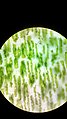
 Onion cells under the fluorescence microscope 1.jpg 2,110 × 2,099; 638 KB
-
 Onion cells under the fluorescence microscope 2.jpg 2,081 × 2,077; 545 KB
-
 Onion cells under the fluorescence microscope 3.jpg 2,101 × 2,159; 581 KB
-
 Onion Cells Under the Microscope.jpg 2,880 × 3,164; 1.91 MB
-
 Onion epidermal cell.jpg 1,602 × 1,771; 938 KB
-
 Onion epidermal cells.jpg 2,448 × 3,264; 424 KB
-
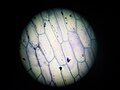
 Onion epidermis high power.jpg 1,024 × 768; 164 KB
-
 Onion flake. Cells. SEM-BSE.jpg 3,072 × 2,304; 3.43 MB
-
 Onion skin cells under a microscope.jpg 960 × 960; 254 KB
-
 Onioncell microscopic photo using foldscope.jpg 1,840 × 3,264; 1.88 MB
-
 OnionCells.jpg 640 × 240; 28 KB
-
 Pecoros 1024x1024.jpg 1,024 × 1,024; 263 KB
-
 Plasmolisi vista al microscopio.jpg 720 × 927; 267 KB
-
Plasmolysis and deplazmoliz onion epidermal cells.ogv 59 s, 768 × 576; 4.34 MB
-
Plazmoliz and deplazmoliz.ogv 21 s, 768 × 576; 1.38 MB
-
 The backbone of the onion!.jpg 1,032 × 774; 76 KB
-
 Κύτταρα κρεμμυδιού.jpg 640 × 480; 240 KB
-
 Клетки Лука ре́пчатого (лат. Állium cépa).jpg 288 × 352; 10 KB
-
 Клетки эпителия репчатого лука. Увеличение в 56 раз.jpg 776 × 777; 141 KB
-
 Клетки эпителия репчатого лука.jpg 4,208 × 3,120; 1.43 MB
-
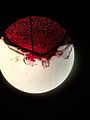
 Красный лук 2.jpg 2,592 × 1,944; 3.84 MB
-
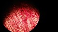
 Красный лук 3.jpg 2,592 × 1,944; 2.94 MB
-
 Кристаллы оксалатов в шелухе лука 1.tif 5,283 × 3,522; 53.26 MB
-
 Кристаллы оксалатов в шелухе лука 2.tif 5,382 × 3,588; 55.28 MB
-
 Кристаллы оксалатов в шелухе лука 3.tif 4,895 × 3,263; 91.42 MB
-
 Кристаллы оксалатов в шелухе лука 4.tif 3,789 × 2,526; 54.79 MB
-
 Кристаллы оксалатов в шелухе лука 5.tif 3,908 × 2,605; 58.28 MB
-
 Лук под микроскопом.jpg 1,536 × 2,048; 382 KB
-
 Мікрофотографія клітин цибулі.jpg 1,000 × 1,000; 266 KB
-
 Шелуха лука с кристаллами оксалата кальция.jpg 3,830 × 2,875; 8.64 MB
-
 Эпидермис лука.jpg 1,600 × 1,200; 731 KB


.jpg)


.png)


.jpg)


